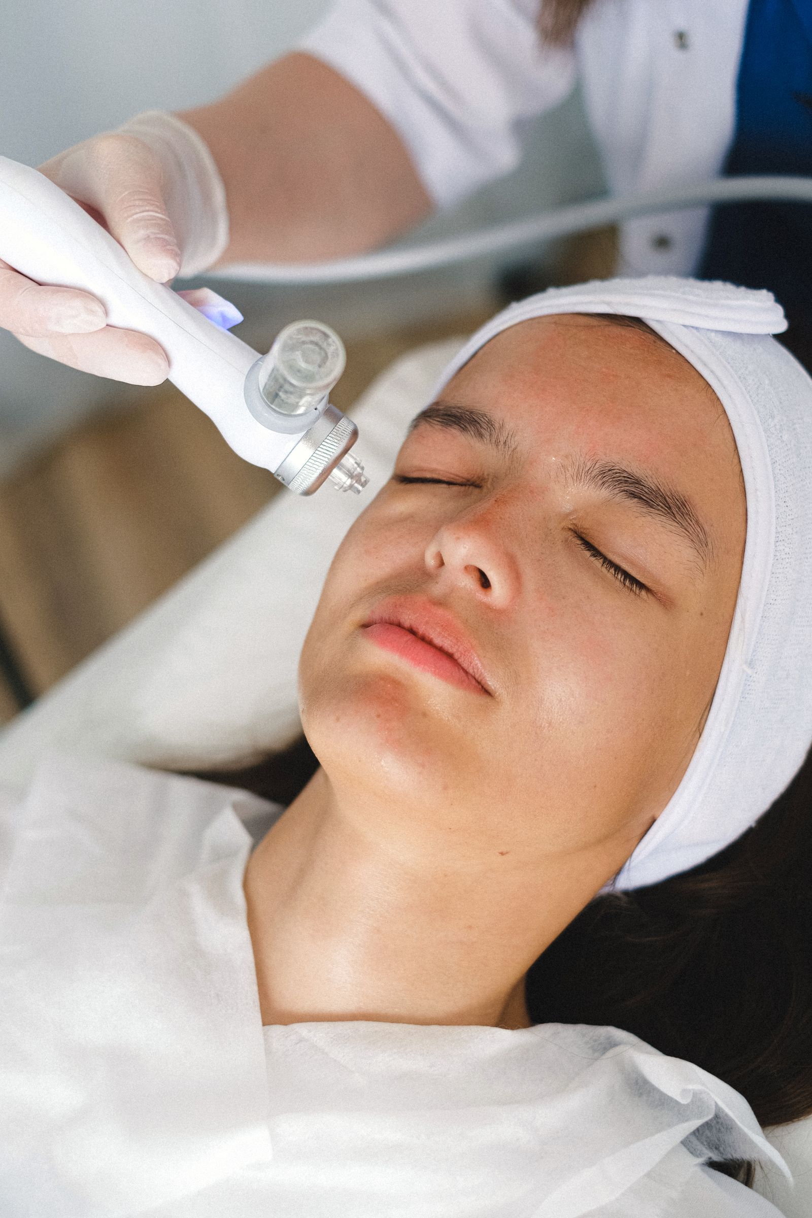
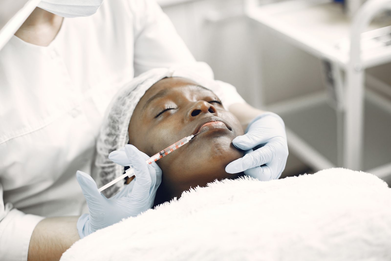
The distinction between PRP and PRF goes far beyond marketing labels. These autologous platelet concentrates behave differently at the cellular level, release growth factors on separate timelines, and occupy distinct clinical niches. We see patients arrive convinced one is universally superior, when the reality is more precise: each therapy wins in specific anatomic and procedural contexts.
How preparation protocols create different biologics
PRP (Platelet-Rich Plasma) and PRF (Platelet-Rich Fibrin) start with the same blood draw but diverge immediately in centrifugation. PRP uses anticoagulant tubes—typically sodium citrate or acid citrate dextrose—and spins at 3000-3500 rpm for 10-12 minutes. This creates a sharp separation: red blood cells at the bottom, acellular plasma at the top, and a thin buffy coat of concentrated platelets in between. We extract that platelet layer, achieving concentrations of 4-6× baseline in most protocols. The American Academy of Orthopaedic Surgeons notes that standardized PRP preparation lacks universal consensus, but citrate-based systems dominate aesthetic use.
PRF eliminates anticoagulant entirely. Blood goes into plain glass tubes and spins at lower speeds—typically 1300-1500 rpm for 8-12 minutes—in a single centrifugation step. Without anticoagulant, the blood begins clotting during the spin, forming a fibrin matrix that traps platelets, leukocytes, and circulating stem cells in a three-dimensional scaffold. The result is a semi-solid clot with platelets suspended throughout, not concentrated in a thin layer. Studies in the Journal of Oral and Maxillofacial Surgery demonstrate that PRF contains growth factors embedded in fibrin, creating a slow-release reservoir.
The absence of anticoagulant in PRF is not a minor detail—it fundamentally alters growth factor kinetics. PRP, once activated with calcium chloride or thrombin, releases 70% of its growth factors within the first hour. PRF releases approximately 10-15% immediately, then continues secreting PDGF, VEGF, and TGF-β over 7-10 days as the fibrin scaffold degrades. This sustained release explains why PRF performs differently in slower-turnover tissues.
When PRP dominates: paired procedures and epidermal targets
PRP excels when we need immediate, high-concentration growth factor delivery to match the trauma timeline of another procedure. Microneedling creates thousands of controlled micro-injuries; combining it with PRP floods those channels with PDGF and EGF exactly when fibroblasts upregulate collagen synthesis. A 2020 study in the Journal of Cosmetic Dermatology found that microneedling plus PRP produced 34% greater collagen density at 90 days compared to microneedling alone, measured by acoustic microscopy.
We also prefer PRP after ablative laser (CO₂, Erbium:YAG) because the immediate growth factor surge accelerates re-epithelialization. Post-laser healing depends on keratinocyte migration from adnexal structures; EGF in PRP directly stimulates that migration. Anecdotally, we see crust shedding 1-2 days earlier with PRP application, and erythema resolving faster. The liquid consistency of PRP allows even distribution across uneven laser-treated surfaces, which semi-solid PRF cannot match.
Scalp applications favor PRP for different reasons. Hair follicles respond to VEGF and hepatocyte growth factor in the active anagen phase; the PRP bolus delivers those factors when follicular papilla cells are metabolically primed. Research published in Dermatologic Surgery showed that PRP injections every 4 weeks for 3 months increased mean hair density by 20-25 hairs/cm² in androgenetic alopecia patients. PRF's slow release doesn't align with the scalp's treatment rhythm.
Where PRF outperforms: volume restoration and anatomic complexity
PRF claims its territory in areas where we need structural scaffold and prolonged growth factor exposure. Under-eye hollowing is the clearest example. The tear trough sits over orbicularis oculi muscle with minimal subcutaneous fat and pronounced vascularity. Injecting liquid PRP here risks rapid absorption and uneven distribution. PRF's fibrin matrix stays in place, providing both immediate (though subtle) volume and a sustained stimulus for neocollagenesis over weeks.
In our clinic, we inject PRF in a multi-plane technique: deeper aliquots along the orbital rim periosteum, superficial threads in the dermal-subdermal junction. The fibrin scaffold integrates into native tissue architecture rather than dissipating. Patients notice progressive improvement over 6-8 weeks—not the immediate plumping of hyaluronic acid, but gradual thickening of the dermal-epidermal junction. Studies in aesthetic medicine journals report that PRF under-eye treatments show durability at 9-12 months, likely because the fibrin matrix stimulates adipocyte differentiation from precursor cells.
PRF also suits nasolabial folds, marionette lines, and other areas where we want volumization without filler but with more than a transient signal. The leukocyte content in PRF—higher than in PRP due to the single-spin protocol—adds antimicrobial peptides and additional growth factors from white blood cells. This makes PRF theoretically safer in areas with higher bacterial colonization, though clinical infection rates with either therapy are vanishingly low when sterile technique is maintained.
Longevity and maintenance intervals differ measurably
PRP typically requires a series of 3 treatments spaced 4-6 weeks apart, then maintenance every 6-9 months. The immediate release and rapid platelet apoptosis mean the biologic signal is intense but brief. We see peak clinical improvement—measured by patient-reported texture and photographic analysis—around 8-12 weeks after the third treatment. After that, collagen remodeling continues passively, but new stimulus is needed to sustain gains.
PRF often follows a different schedule: 2-3 treatments spaced 6-8 weeks apart, with maintenance stretched to 10-14 months. The prolonged growth factor release and fibrin scaffold persistence create a longer active phase. Clinical observations published in aesthetic surgery literature suggest that PRF's leukocyte content may extend anti-inflammatory signaling, potentially contributing to longer-lasting dermal remodeling. However, head-to-head longevity studies are sparse, and most comparative data comes from dental and orthopedic fields, not aesthetic medicine.
Cost per treatment is comparable—both require the same blood draw volume (15-30 mL depending on protocol)—but total cost over 18 months may favor PRF due to fewer sessions. That said, PRP's versatility in combination treatments often makes it the more frequently deployed option in a comprehensive aesthetic protocol.
Technical variables that change outcomes more than PRP-vs-PRF choice
The device and spin protocol matter more than practitioners admit. PRP systems range from simple manual centrifuges to closed automated systems like Emcyte PurePRP or Arthrex Angel. Automated systems reduce operator variability but cost significantly more. PRF relies on precise timing: spin too short, and platelet concentration drops; spin too long, and the clot becomes too dense to inject smoothly. We use the IntraSpin system for PRF, which standardizes tube position and deceleration curves.
Activation method for PRP influences release kinetics. Calcium chloride is most common, but some protocols use thrombin or mechanical activation (freeze-thaw cycles). Research in the Journal of Tissue Engineering shows that thrombin activation releases growth factors more completely than calcium alone, but it also accelerates platelet aggregation, which can clog fine needles during injection. We use calcium chloride for most PRP applications because it balances activation speed with injectability.
Injection depth and volume per pass are critical for both. Intradermal PRP (0.05-0.1 mL per wheal) targets photoaging and texture. Deep dermal to subcutaneous PRF (0.2-0.3 mL per depot) addresses volume loss. Cross these wires—inject PRP too deep or PRF too superficial—and you lose the anatomic advantage of each therapy. Needle gauge matters: we use 30G for PRP, 27G for PRF due to viscosity. Cannulas (25G blunt-tip) work well for PRF in the midface, reducing bruising in vascular zones.
When to combine them or choose neither
Some protocols layer PRP and PRF in the same session. A typical sequence: deep PRF for structure, followed by superficial PRP for immediate epidermal signaling. This is common in combined tear trough and lower lid skin rejuvenation, where we want both volume scaffolding (PRF) and surface texture improvement (PRP). Anecdotal reports in aesthetic medicine forums suggest additive effects, but no robust studies quantify the benefit over monotherapy.
Neither PRP nor PRF is appropriate when patients want immediate, dramatic correction. A 50-year-old with 2 mL of volume loss per midface will not achieve sufficient correction with autologous therapies alone. Here, hyaluronic acid filler provides the structural restoration, and PRP or PRF become adjuncts to improve skin quality around the filler. We also avoid platelet therapies in active infection, uncontrolled diabetes (impairs platelet function), or patients on antiplatelet drugs (aspirin, clopidogrel) that blunt growth factor release.
Patients with unrealistic timelines—wanting full correction in 2 weeks—are better served by other modalities. Platelet therapies are fundamentally regenerative, not restorative. They amplify the body's repair capacity but cannot override biologic limits. Setting this expectation upfront prevents disappointment and ensures patients select the therapy aligned with their goals and patience.